
Are all lung cancers the same? NOT EVEN CLOSE! – The scan shows where. The genome shows what
Every November, Lung Cancer Awareness Month asks us to look harder at the world’s deadliest cancer. Why does awareness still matter? The numbers answer that.
The Scale of the Problem
Lung cancer was the most commonly diagnosed cancer in the world in 2022, with about 2.5 million new cases, almost one in eight of all new cancer cases (IARC). It was also the leading cause of cancer death, with about 1.8 million deaths that year (WHO).India carries a heavy share of that burden. IARC’s latest country estimates for India (
GLOBOCAN, 2024 estimates) count 112,659 new lung cancer cases, making it the third most common cancer, and 98,687 deaths, making it the leading cause of cancer death at 10.9% of all cancer deaths (IARC India fact sheet).
Put those two Indian numbers side by side. Deaths come to roughly 88 for every 100 new cases, a gap that reflects, among other things, how often the disease is found late.
Why Lung Cancer Slips Through
If the numbers are this stark, why is the disease still found so late? Three reasons stand out.
- It starts quietly. Early symptoms are mild and easy to dismiss as ordinary respiratory problems, so many patients are diagnosed at an advanced stage, when treatment options are limited (WHO).
- It looks like other things. In India, where tuberculosis is also common, a persistent cough is often assumed to be an infection or TB first, which can delay the right investigation.
- It does not only affect smokers. Tobacco remains the leading cause, accounting for 60 to 70% of preventable lung cancers, but air pollution, second-hand smoke, occupational exposures and radon also contribute (WHO). A person who has never smoked may not suspect lung cancer, and may be assessed late.
Screening high-risk people can help, and WHO notes that it has reduced mortality in well-resourced health systems. But a scan only finds a tumour. It does not tell you what kind of tumour it is, and that is where our story turns.
Multiple Patients, One Diagnosis
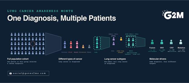
Picture four patients with the same diagnosis: stage IV lung adenocarcinoma. Their CT scans show similar disease. Their biopsies look identical under the microscope. As far as the scan and the slide can tell, they are the same case.
They are not. Each tumour is driven by a different kind of molecular change.
- A fusion. In the first patient, the ALK gene has fused with a partner gene, producing a hybrid protein that keeps telling the cell to grow.
- A mutation. In the second, a short stretch of the EGFR gene has been deleted, leaving its growth signal permanently switched on.
- A single nucleotide variant (SNV). In the third, a single letter of the KRAS gene’s code has changed, swapping one amino acid for another. This change is known as G12C.
- A copy number change. In the fourth, the tumour carries extra copies of a gene such as MET, so a growth signal is produced in excess. Clinicians increasingly look for this change, particularly when a tumour stops responding to earlier treatment.
Each is a different engine of growth, and each can point to a different treatment decision. The scan could not see any of them. The slide could not see any of them. Only a molecular test could, and only a test built to look for that kind of change: a test designed for point mutations will not reveal a fusion or a copy number change.
Now change one detail. For any of these four patients, the test is never run, or it is run on a method that cannot see that kind of alteration and returns a clean result. Nothing about the scan or the slide looks wrong, so no one has a reason to question it. The right option may never be considered.
(These are illustrative scenarios, not real patients, but they play out in clinics every day.)
That is the quiet gap behind the statistics above. Awareness is not only about spotting symptoms sooner. It is about making sure that once lung cancer is found, the tumour is also understood.
What the Molecular Layer Reveals
Imaging shows where a tumour is, and histology shows what it resembles. Molecular testing shows what is driving it. In many non-small cell lung cancers, one altered gene acts as the engine of growth, and identifying it can change how the patient is managed. Each class of alteration, however, presents its own detection challenge.
| Alteration | Type of change | Detection challenge | Best-suited method |
| EGFR (exon 19 deletions, L858R, exon 20 insertions) | SNV / indel | Many variants across several exons | RT-PCR for common hotspots; NGS for rarer variants |
| KRAS (including G12C) | SNV | Multiple hotspot codons | RT-PCR or NGS |
| BRAF V600E | SNV | Single hotspot, but low frequency | RT-PCR or NGS |
| ALK, ROS1, RET, NTRK | Gene fusion | Variable breakpoints that DNA-only assays can miss | RNA-based NGS; RT-PCR for known fusion variants such as EML4-ALK |
| MET exon 14 skipping | Splice-altering variants | Many different DNA changes lead to the same event | RNA-based or broad DNA NGS |
| HER2 (ERBB2) mutations | SNV / indel | Spread across the gene | NGS |
| Tumour mutational burden (TMB) | Genome-wide measure | Needs adequate panel size and depth | NGS |
Every alteration above except TMB is an actionable biomarker that guides targeted therapy selection, while TMB informs immunotherapy decisions. That is why detecting each one accurately matters.
The frequency of these alterations also depends on the patient population. EGFR mutations, for example, are found in a considerably larger share of lung adenocarcinomas in Asian populations than in Western ones, which is one reason molecular testing carries particular weight in India.
Finding the Answer Earlier: Genes2Me Solutions
At Genes2Me, we build molecular diagnostics around these detection challenges, so the genetic answer arrives early in the diagnostic pathway rather than late or not at all. Our lung cancer portfolio matches the method to the sample and the clinical question, across three situations a laboratory faces every day.
- When the tumour sample is available: the FOCUS Lung NGS Assay profiles 73 DNA genes and 18 RNA fusion genes from FFPE or fresh tissue in a workflow of about 1.5 days, covering SNVs, indels, CNVs, fusions, TMB and MET exon 14 skipping.
- When tissue is limited or a biopsy is difficult: the ctDNA Lung panel in our liquid biopsy portfolio profiles 32 genes from a blood or plasma sample, using as little as 10 to 20 ng of cell-free DNA.
- When a fast answer on the key drivers is needed: our CE-IVD oncology RT-PCR kits, including EGFR-Q, KRAS-Q, BRAF-Q and EML4-ALK, run on widely used real-time PCR instruments.
These tools work on diagnosed disease, helping laboratories and clinicians learn what is driving a tumour as early as possible.
Awareness That Changes the Outcome
Awareness alone will not make lung cancer less common. But what happens after the first symptom, the first scan and the first diagnosis can change.
For the public, that means taking a cough that will not clear, or breathlessness that does not settle, seriously, and getting it properly investigated, including in people who have never smoked. For anyone facing a diagnosis, it means asking one more question: has the tumour itself been tested for the genetic alterations that could guide treatment? For clinicians and laboratories, it means making that testing a routine part of the pathway, not an afterthought.
A scan can show where a tumour is. What is driving it is written in its genome, and every patient with lung cancer deserves to have it read.
