
Decoding Lung Cancer: Precision Genomics with FOCUS Lung & CliSeq
A tumour sample may look like a small piece of tissue, but from a genomic point of view, it contains a large amount of information. In lung cancer, that information can be especially important because tumours can have different genetic changes. Mutations, fusions, and biomarkers can differ between tumours, and these differences can affect both the progression of the disease and its response to treatment.
The challenge is not simply finding these changes. Once a sample is sequenced, the result is a large collection of data. That data has to be checked and interpreted before it can provide useful information.
The FOCUS Lung NGS Assay from Genes2Me is designed for the testing part of this process. CliSeq Interpreter takes the sequencing information and carries out the bioinformatics analysis. When the two are viewed together, the process becomes a connected path from the original tissue sample to genomic findings and reports.
The genetic information being examined
FOCUS Lung covers 73 DNA genes and 18 RNA fusions. The panel includes guideline-recommended targets such as EGFR, KRAS, ALK, ROS1, RET, and NTRK.
The assay has a 261 Kb target size and covers whole coding sequences. It can detect several types of genomic changes, including SNVs, indels, CNVs, and fusions. It also looks at biomarkers such as tumour mutational burden, commonly referred to as TMB, and MET exon 14 skipping.
The reported performance figures for the assay are:
> 99% coverage uniformity
97.2% reproducibility
75–84% on-target ratio
98.6% sensitivity at 1% VAF
These numbers are useful when considering how the assay performs across different samples. In particular, the sensitivity at 1% VAF matters when a variant is present at a low level. A tumour sample can contain different groups of cells, and a genetic change may therefore occur in only a small proportion of the sample.
Getting the sample ready
The workflow starts before the sequencing machine is involved.
The sample can come from FFPE or fresh tissue. It first goes through extraction so that the material required for testing can be obtained.
Next comes library preparation. Hybrid-capture enrichment is used at this stage. Once the library is prepared, it can move to sequencing.
FOCUS Lung supports sequencing across Illumina, MGI, Thermo Fisher, or Element Biosciences platforms.
After sequencing, the sample itself is no longer the main focus. The large amount of information generated from it becomes the next thing that needs to be handled. This is where CliSeq Interpreter comes in.
Turning sequencing data into variants
Millions of reads can come from a sequencing run. At this stage, simply having the data is not enough. The reads have to be checked and processed before the relevant genetic findings can be identified.
CliSeq Interpreter is a browser-based bioinformatics platform used for this work.
The analysis begins with quality control and trimming. These steps help ensure that the reads meet high-fidelity standards.
The reads are then aligned to the GRCh38 reference genome using BWA. Once the alignment is completed, somatic variant calling is carried out using GATK Mutect2. The process is optimized for SNVs, indels, CNVs, and fusions.
The variants identified at this stage are then annotated. Information from databases including ClinVar, dbSNP, gnomAD, and 1000 Genomes is used for annotation.
There is still another step before the results can be considered in a focused way.
Filtering the findings
A sequencing result can contain many variants. That does not mean every variant has the same importance.
CliSeq Interpreter applies filtering using factors such as allele frequency, pathogenicity, depth, and phenotype relevance. This helps separate findings according to their relevance rather than treating every variant in the same way.
This part of the workflow is important because genomic analysis is not just about producing a long list of mutations. The information needs to be narrowed down so that biologically and clinically significant findings can be given more attention.
The result is a more focused set of genomic findings instead of an unorganised collection of sequencing data.
How the findings are reported
Once the analysis is finished, the results have to be presented in a way that researchers can actually work with.
CliSeq Interpreter provides a CSM Report. This report gives a consolidated summary of clinically significant mutations, with the findings annotated and filtered for clarity.
The Data Quality Report is different. It contains detailed QC metrics that help validate the sequencing and analysis performance.
There is also a Raw VCF. This contains the complete variant calls in standard format and allows computational re-analysis.
The Annotated VCF contains functional and database annotations and can be used for downstream research.
The reports are available through a single dashboard. From there, researchers can follow the analysis status, look at QC checkpoints, and download the required outputs.
So the dashboard is not just the final destination for the report. It also provides a place to follow what has happened during the analysis.
From genetic findings to treatment options
Genomic testing becomes more meaningful when the findings can be considered alongside treatment options.
For example, an ALK fusion can be connected with Alectinib, Brigatinib, and Lorlatinib.
RET fusions can be connected with Pralsetinib and Selpercatinib.
For ROS1 fusions, the examples include Crizotinib, Entrectinib, and Repotrectinib.
These are FDA-approved therapies associated with the respective genomic findings. This connection is an important part of precision oncology because it shows how information about the tumour can be related to potential therapeutic options.
Looking at the workflow as one process
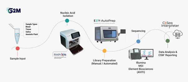
The entire journey can be reduced to six stages:
Sample → Library Preparation → Sequencing → Analysis → Reporting → Therapy Connection
The first stage involves FFPE or fresh tissue. After extraction and library preparation, the sample is sequenced. The resulting data then enters CliSeq Interpreter.
From there, it goes through quality control and trimming, alignment to GRCh38 using BWA, somatic variant calling with GATK Mutect2, annotation, and filtering.
The findings are then available through the different reports.
Seen this way, the assay and the software are doing different jobs, but the jobs are connected. The FOCUS Lung NGS Assay provides the molecular testing, while CliSeq Interpreter helps turn the resulting sequencing information into something that can be examined and reported.
Why this matters specifically for lung cancer
The genetic profile of a lung tumour is not necessarily the same as that of another lung tumour. Different mutations, fusions, and biomarkers may be present. These differences can have an effect on disease progression and therapeutic response.
That is why molecular information can add another level of understanding to lung cancer.
Instead of stopping at the diagnosis, genomic testing allows the tumour to be examined for specific genetic characteristics. The FOCUS Lung NGS Assay provides that testing capability, and CliSeq Interpreter takes the resulting information through the analysis and reporting stages.
The value is therefore in the complete process rather than in one individual step.
Final thoughts
What initially looks like a complicated collection of laboratory and computer-based steps starts to make more sense when the workflow is followed from beginning to end.
A tissue sample is extracted. It is prepared and sequenced. The sequencing produces millions of reads. Those reads are checked, aligned, and analyzed. Variants are identified, annotated, and filtered. The important findings are then organized into reports.
The FOCUS Lung NGS Assay and CliSeq Interpreter bring these stages together for lung cancer genomic analysis.
There is a lot of technical detail involved, from 73 DNA genes and 18 RNA fusions to GRCh38, BWA, GATK Mutect2, ClinVar, VCF files, and QC metrics. But the purpose behind all of these steps is fairly straightforward: to understand the genetic characteristics of the tumour in greater detail.
When that information is connected with relevant therapeutic options, genomic testing becomes more than a way of collecting genetic data. It becomes a way of adding molecular information to the understanding of lung cancer.
One sample. One workflow. A clearer tomorrow.
